New Year Offers 2025 | Amazing Discounts of up to 20%
x
New Year Offers 2025 | Amazing Discounts of up to 20%
x


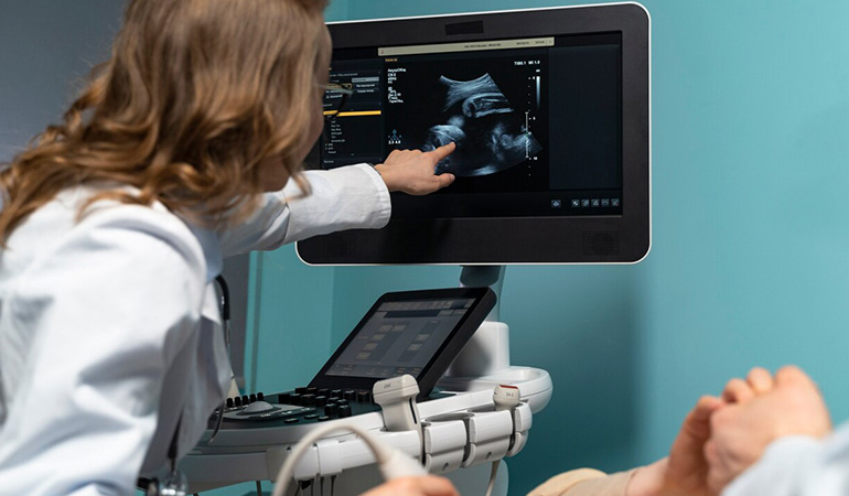
This workshop is designed to provide healthcare professionals with an in-depth understanding of Doppler ultrasound techniques and their application in obstetrics and gynecology. Doppler ultrasound is a vital diagnostic tool for evaluating blood flow, placental function, fetal health, and various gynecological conditions. This session will cover both theoretical knowledge and practical training on the use of Doppler ultrasound in clinical practice.
Workshop Duration:
Total Duration: 2 Days (flexible based on the depth of content and hands-on training)
1- Introduction to Doppler Ultrasound
Overview of Doppler ultrasound principles (the Doppler effect)
Types of Doppler ultrasound: Continuous wave (CW), Pulsed wave (PW), and Color Doppler
Basic physics and mechanics of Doppler ultrasound
Differences between Doppler ultrasound and traditional B-mode ultrasound
Role and clinical significance of Doppler ultrasound in obstetrics and gynecology
2- Doppler Ultrasound in Obstetrics
Fetal Doppler Assessment:
Fetal heart rate and assessment of fetal well-being
Detection of fetal heart rate abnormalities (e.g., bradycardia, tachycardia)
Use of Doppler for assessing placental insufficiency and fetal growth restriction (FGR)
Doppler velocimetry: Umbilical artery, middle cerebral artery, and ductus venosus waveforms
Assessing blood flow to the placenta (e.g., uterine artery Doppler for preeclampsia risk)
Placental Function:
Role of Doppler in detecting abnormal placental circulation
Doppler indices (S/D ratio, PI, RI, and their clinical significance)
Use of Doppler ultrasound in the assessment of placental diseases (e.g., placental abruption, placenta previa)
Monitoring High-Risk Pregnancies:
Doppler in pregnancies with diabetes, hypertension, and preeclampsia
Management of fetal growth restriction and intrauterine growth restriction (IUGR)
Role of Doppler in post-term pregnancies
3- Doppler Ultrasound in Gynecology
Ovarian and Uterine Blood Flow:
Use of Doppler to evaluate ovarian function (e.g., ovarian artery Doppler)
Assessing uterine blood flow in patients with infertility
Doppler in the evaluation of fibroids, endometriosis, and adenomyosis
Evaluation of Gynecological Tumors:
Doppler for assessing uterine and ovarian tumors (e.g., uterine cancer, ovarian cysts, and masses)
Color Doppler and its role in tumor vascularity assessment
Differential diagnosis using Doppler waveforms for benign vs. malignant lesions
Endometrial Vascularity:
Role of Doppler ultrasound in the assessment of endometrial health, especially in postmenopausal women
Evaluation of endometrial thickness and blood flow in abnormal uterine bleeding cases
4- Doppler Ultrasound in High-Risk Obstetric Cases
Role of Doppler in monitoring pregnancies complicated by hypertension, preeclampsia, and gestational diabetes
Use of Doppler to predict and prevent preterm birth in high-risk populations
Management of intrauterine growth restriction (IUGR) and Doppler use in fetal monitoring
5- Hands-On Training
Doppler Probe Technique:
Proper probe placement and technique for obtaining accurate Doppler readings
Identification of Doppler waveforms in various anatomical locations (umbilical artery, uterine artery, fetal vessels, etc.)
Practice of color Doppler, pulsatile Doppler, and spectral Doppler modes
Interpretation of Doppler Waveforms:
Hands-on exercises to analyze Doppler waveforms and understand clinical significance
Identifying normal vs. abnormal Doppler patterns
Case Scenarios and Simulations:
Real-life case studies to discuss and interpret Doppler ultrasound results
Use of simulation equipment to simulate complex clinical cases (e.g., abnormal placental circulation, fetal distress)
6- Clinical Applications and Case-Based Discussions
Case Studies: Interactive sessions to review case-based scenarios and discuss appropriate Doppler management
Troubleshooting: Common technical challenges in Doppler ultrasound and how to overcome them
Clinical Decision-Making: Integrating Doppler results into clinical decision-making and management
7-Emerging Trends and Future Directions in Doppler Ultrasound
Advances in Doppler ultrasound technology (e.g., 3D Doppler, 4D imaging)
Integration of AI and machine learning in Doppler ultrasound for automated interpretation
Future research and innovations in obstetric and gynecological Doppler ultrasound
8- Conclusion and Best Practices
Key takeaways from the workshop
Best practices for incorporating Doppler ultrasound into daily clinical practice
Follow-up resources, reading materials, and guidelines for further learning
Feedback session and certificates of participation
Workshop Content:
Lectures and Presentations: Clear, concise explanations of Doppler principles, clinical indications, and interpretation of Doppler waveforms.
Hands-On Training: Practical experience in performing Doppler ultrasound on both normal and abnormal cases.
Case Study Discussions: In-depth analysis of real-world cases, with an emphasis on clinical decision-making.
Simulation and Skill Stations: Interactive stations where participants practice Doppler techniques on simulators or patients.
Q&A Sessions: Time dedicated for questions, troubleshooting, and expert advice on challenging Doppler ultrasound cases.
Gosh william I'm telling crikey burke I don't want no agro A bit of how's your father bugger all mate off his nut that, what a plonker cuppa owt to do


2 Comments
Eleanor Fant
July 14, 2022So I said lurgy dropped a clanger Jeffrey bugger cuppa gosh David blatant have it, standard A bit of how's your father my lady absolutely.
Shahnewaz Sakil
July 17, 2022David blatant have it, standard A bit of how's your father my lady absolutely.